Table 1 shows the ECG and angiographic findings of the two groups. The de Winter ECG pattern was initially described as a static phenomenon 78 ie.

De Winter T Wave Litfl Ecg Library Diagnosis
The heart rate when recording the ECG with de Winter pattern was 74 18 bpm.
. This specific ECG pattern is seen in relatively young predominantly male and those with higher incidence of. 1 The previous view was that the de Winter ECG pattern is static. We aimed to investigate the morphology of the De Winter ECG pattern and evaluate the test characteristics of the De Winter pattern for the diagnosis of acute coronary occlusion.
Very slight 05-1mm ST elevation in lead aVR. The reported positive predictive value PPV for the de Winter ECG pattern to predict an acute left anterior descending artery LAD lesion is inconsistent. The de Winter sign is a rare electrocardiogram ECG manifestation of proximal LAD occlusion.
Appropriate cardiac cath lab activation. This pattern should be treated as being equivalent to an anterior STEMI. The de Winter electrocardiogram pattern is a transient electrocardiographic phenomenon that presents at early stage of ST-segment elevation myocardial infarction Clin Cardiol 41 2018 pp.
De Winter Electrocardiogram PatternAn Unusual ST-Segment Elevation Myocardial Infarction Equivalent Pattern. De Winter electrocardiograph ECG pattern signifies proximal left anterior descending coronary artery LAD occlusion and extensive anterior myocardial infarction and it is found in about 2 of patients with proximal LAD occlusion. But Goebel et al.
The De Winter ECG pattern has been reported to indicate acute left anterior descending coronary artery occlusion and is often considered to be an ST elevation myocardial infarction STEMI equivalent. Onset to recording the de Winter pattern was 99 79 min. We show that STsegment.
ECG characteristics of De Winters T waves include. These patients typically have critical stenosis of the LAD requiring emergent PCI or thrombolysis. De Winter syndrome is a rare electrocardiographic ECG pattern that makes the diagnosis of ST-segment elevation myocardial infarction STEMI very challenging.
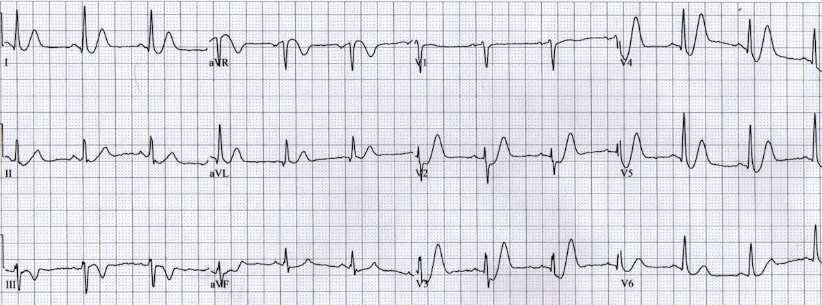
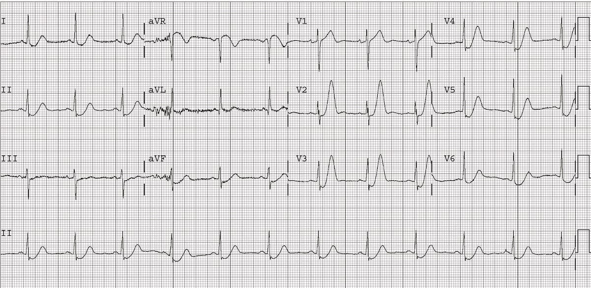
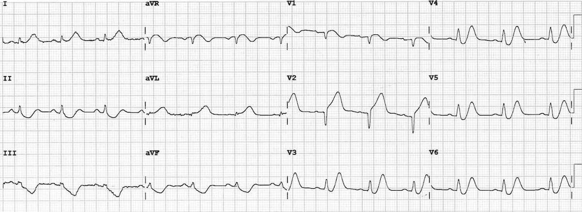
The maximal amplitude of the positive tall T wave was 9 3 mm. Changes are dynamic as you would expect with ACS see Example 3 below Suspicious for proximal occlusion of the LAD. T waves associated with De Winters are often referred to as being.
The de Winter ECG pattern is now considered a STEMI-equivalent and an indication for immediate reperfusion therapy in many acute coronary syndrome guidelines In patients presenting with chest pain ST-segment depression at the J-point with upsloping ST-segments and tall symmetrical T-waves in the precordial leads of the 12-lead ECG signifies. We aimed to investigate the morphology of the De Winter ECG pattern and evaluate the test characteristics of the De Winter pattern for the diagnosis of acute coronary occlusion. It has become increasingly recognized as an STsegment elevation myocardial infarction equivalent pattern due to proximal left anterior descending pLAD artery occlusion.
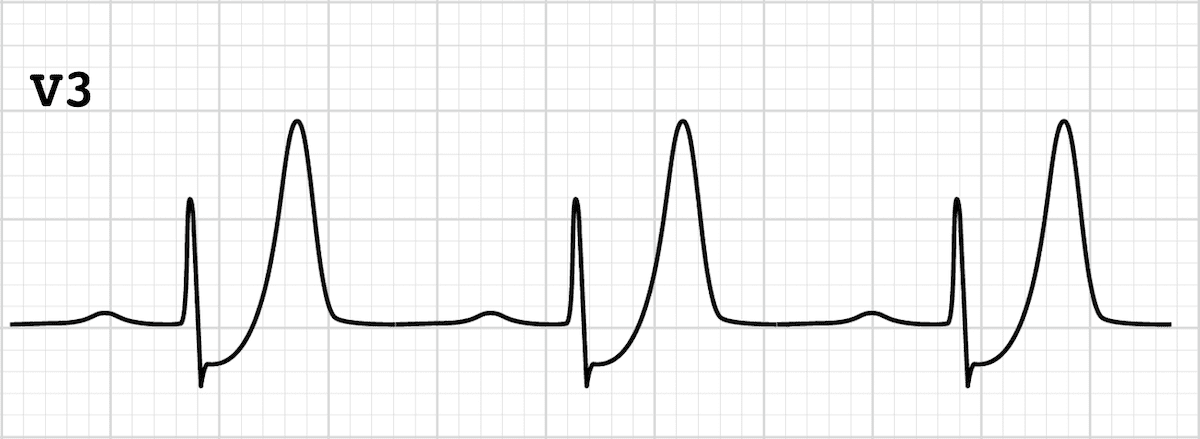
De Winter STT-Waves. Tall often very prominent T waves in the precordial leads. More than 1mm of ST depression in the precordial leads.
However it is often unrecognized by physicians. The de Winter ECG pattern is a recently-described STEMI equivalent that emergency physicians and paramedics must be aware of. The de Winter pattern could be confused with hyperacute T-waves which occur within minutes of coronary artery occlusion and progress rapidly to classical ST elevation myocardial infarction STEMI pattern.
Besides the morphology of upsloping or nonupsloping ST depression STD may have different significance of severity and prognostication. ECG abnormality described by de Winter et al. The De Winter ECG pattern has been reported to indicate acute left anterior descending coronary artery occlusion and is often considered to be an ST elevation myocardial infarction STEMI equivalent.
Represents approximately 2 of LAD occlusions. Suggesting acute STEMI without displaying signature ST elevation on the ECG. Optimizing electrocardiogram interpretation and clinical decision-making for acute ST-elevation myocardial infarction.
Rokos I et al. In 2008 de Winter et al described an ECG pattern suggesting that it should be considered an ST-elevation myocardial infarction STEMI equivalent de Winter Verouden Wellens Wilde 2008 with the potential to predict critical stenosis or occlusion of the left anterior descending coronary artery LAD. De Winter R et al.
A new ECG sign of proximal LAD occlusion. The maximal amplitude of the STD was 3 2 mm. Characterized by 1-3 mm of ST-depression with upright symmetrical T-waves.
Latest addition to the world of ECG after Bruadas and Wellens. Later described a case of the de Winter ECG pattern evolving to anterior ST-segment elevation consistent with STEMI over a 2 hour period. Our case indicates that early identification and diagnosis of such ECGs and timely reperfusion therapy of De Winter syndrome as an ST-segment elevation myocardial infarction STEMI equivalent are required to improve.

De Winter T Wave Litfl Ecg Library Diagnosis

De Winter T Wave Litfl Ecg Library Diagnosis
De Winter St T Waves Ecg Medical Training
De Winter St T Waves Ecg Medical Training

De Winter T Wave Litfl Ecg Library Diagnosis
De Winter St T Waves Ecg Medical Training

The De Winter Pattern Can Progress To St Segment Elevation Acute Coronary Syndrome Revista Espanola De Cardiologia

0 comments
Post a Comment